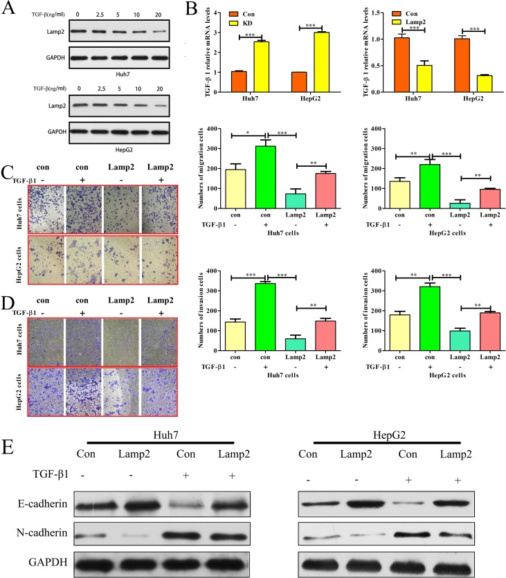
Figure 6. Lamp2 inhibits TGF-β-induced epithelial-mesenchymal transition.
(A) Lamp2 expression levels in Huh7 and HepG2 cells treated with different TGF-β1doses. (B) TGF-β1 mRNA levels were determined using real-time PCR in HCC cells treated with si-NC, siRNA-Lamp2, empty vector or LV-Lamp2. The error bar represents the mean ± SD of triplicate assays. (*p < 0.05, **p < 0.01, and ***p < 0.001; p-values were calculated using Student's t-test). Relative changes in the number of Huh7 and HepG2 cells that migrated (C) and invaded (D) after TGF-β1 treatment or Lamp2 overexpression. (*p < 0.05, **p < 0.01, and ***p < 0.001; p-values were calculated using Student's t-test). (E). Western blot analysis of Lamp2, E-cadherin and N-cadherin expression after TGF-β1 treatment (20 ng/ml) in Huh7 and HepG2 cells that were infected with the LV-NC or LV-Lamp2 lentivirus.

