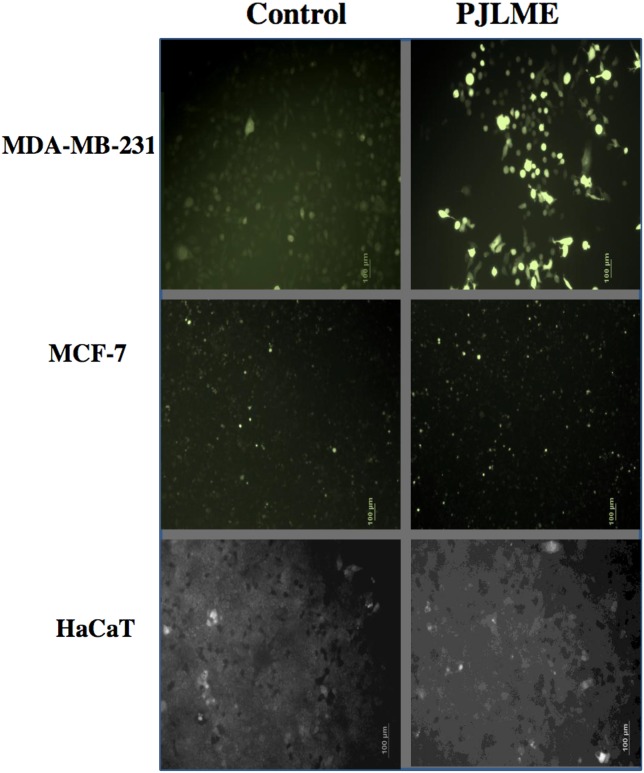
Figure 5. Detection of PJLME induced intracellular reactive oxygen species (ROS).
For detection of ROS, the cells were treated with PJLME (16.8 μg/mL) for 72h and were subsequently exposed to DCFH-DA (10 μM) dye. The results were compared with control set (DMSO 0.1%). Intracellular ROS generation was observed and images were captured by using fluorescence microscope (Olympus, Tokyo, Japan).

