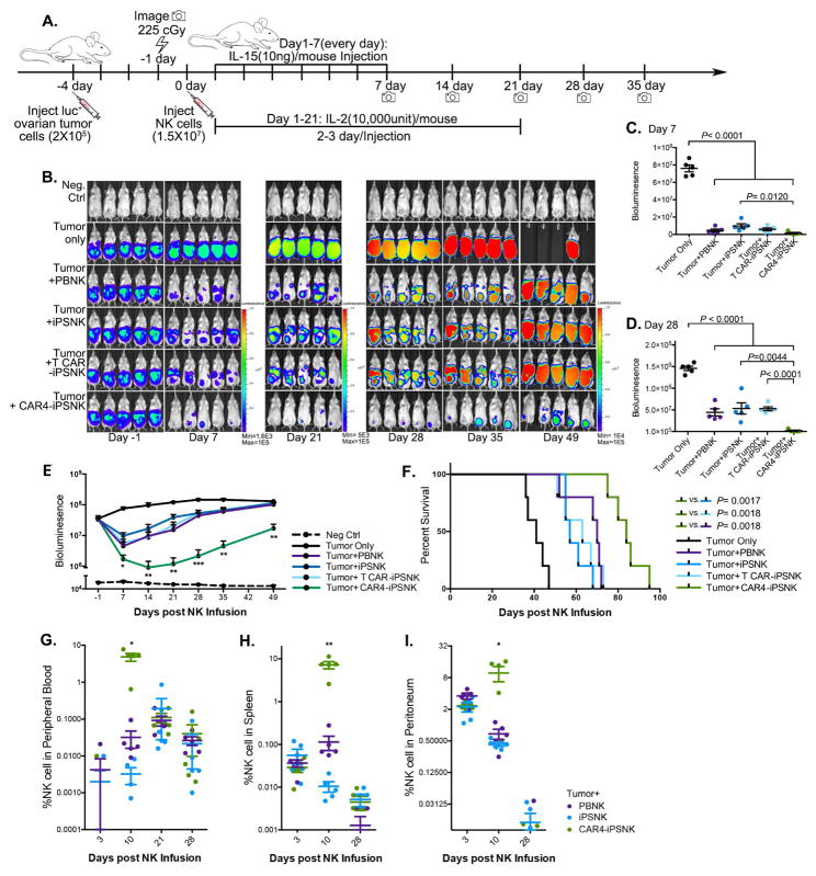
Figure 4. CAR4-expressing iPSC derived NK cells display superior anti-ovarian cancer activity in vivo.
(A) Schematic of in vivo studies using luciferase (luc)-expressing mesohigh A1847 cells in a mouse xenograft model treated with PB-NK cells and iPSC-NK populations and cytokine administration. (B) Tumor burden was determined by weekly bioluminescent imaging (BLI). (C and D) Images of representative time points are shown. Quantification of tumor burden on (C) day 7, and (D) day 28 was plotted based on BLI total flux (photons/sec). (E) Tumor burden of each group was monitored for 49 days post NK population infusion. The BLI data is plotted, mean ± S.D are shown. Statistic: two-tailed Student t-test, T-CAR(meso)-iPSC-NK vs. CAR4(meso)-iPSC-NK. (F) Kaplan-Meier curve representing the percent survival of the experimental groups: Tumor only, or treated with PB-NK cells, iPSC-NK cells, T-CAR(meso)-iPSC-NK cells, or CAR4(meso)-iPSC-NK cells. Statistic: two-tailed Log-rank test. (G–I) Flow cytometric quantification of CD45+CD56+CD3- NK cell population from (G) peripheral blood, (H) spleen, and (I) peritoneal fluid. Each dot represents one recipient mouse. Median ± S.D. is shown.
Statistical analysis by two-tailed one-way ANOVA. *P<0.05, ** P<0.01, *** P<0.001.
See also Figure S7A.

