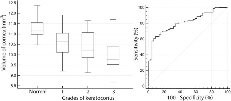
FIGURE 4.

Analyses of volume of cornea as a diagnostic index to detect keratoconus. (Left) Box-whisker plot of central 5 mm volume of cornea in normal eyes and eyes with grades (1, 2, 3) of keratoconus. (Right) Receiver operating characteristic curve of central 5 mm volume of cornea plotted at left.
