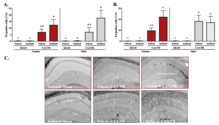
Figure 7.
Neurodegeneration, as indicated by fluoro-jade c positive staining, was increased in both female and male mice at 96 hours post-ischemia. Among females and males, cell death in the CA1 region of the hippocampus was increased in the isolated CA/CPR group relative to sham controls, while the paired CA/CPR group exhibited an intermediate phenotype comparable to both the shams and isolated CA/CPR group (A). This pattern was also present for the CA3 region among females, however, among the males, amelioration of ischemia-induced neurodegeneration from the social environment was not detected (B). A representative image of fluoro-jade c positive staining is presented for each group (C). Scale bar in panel 3C 500 μm.

