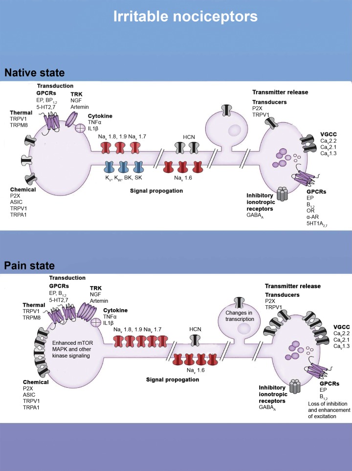
Figure 3.
The irritable nociceptor, molecular mechanisms. The diagram shows channels and modulatory proteins involved in pain transduction, signal propagation, and transmitter release in the spinal dorsal horn. The top panel shows these in the normal state, and the bottom panel shows how changes in these proteins can lead to an irritable nociceptor phenotype. Such changes include increased expression or activity in transducers like TRPV1 or P2X channels, increases in G-protein coupled receptor (GPCRs) like EP receptors, and enhanced signaling in nociceptor terminals. Increases in the expression of voltage-gated sodium channels (Navs) and decreased expression of potassium channels can also shift the balance toward excitation in these nociceptors. Finally, changes in expression of inhibitory and excitatory proteins in the central terminals of nociceptors can also enhance the irritability of these cells. CNS = central nervous system.

