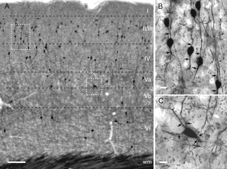
Figure 1.
Light microscopic characterization of YFP-immunolabeled cells in the barrel cortex of the VIPcre-YFP mouse. (A) Distribution of the population of VIP cells in mouse barrel field shown in a 50 μm-thick, osmium-intensified and resin-embedded section. Most cells are located in superficial layers I–IV whereas much fewer are found in deep layers Va, Vb and VI. (B) Morphology of a cluster of bipolar VIP cells in layer II/III (left inset in A). (C) Multipolar VIP cell in layer Va (right inset in A). Arrows indicate primary dendrites. Scale bars = 100 μm in A; 10 μm in B, C.

