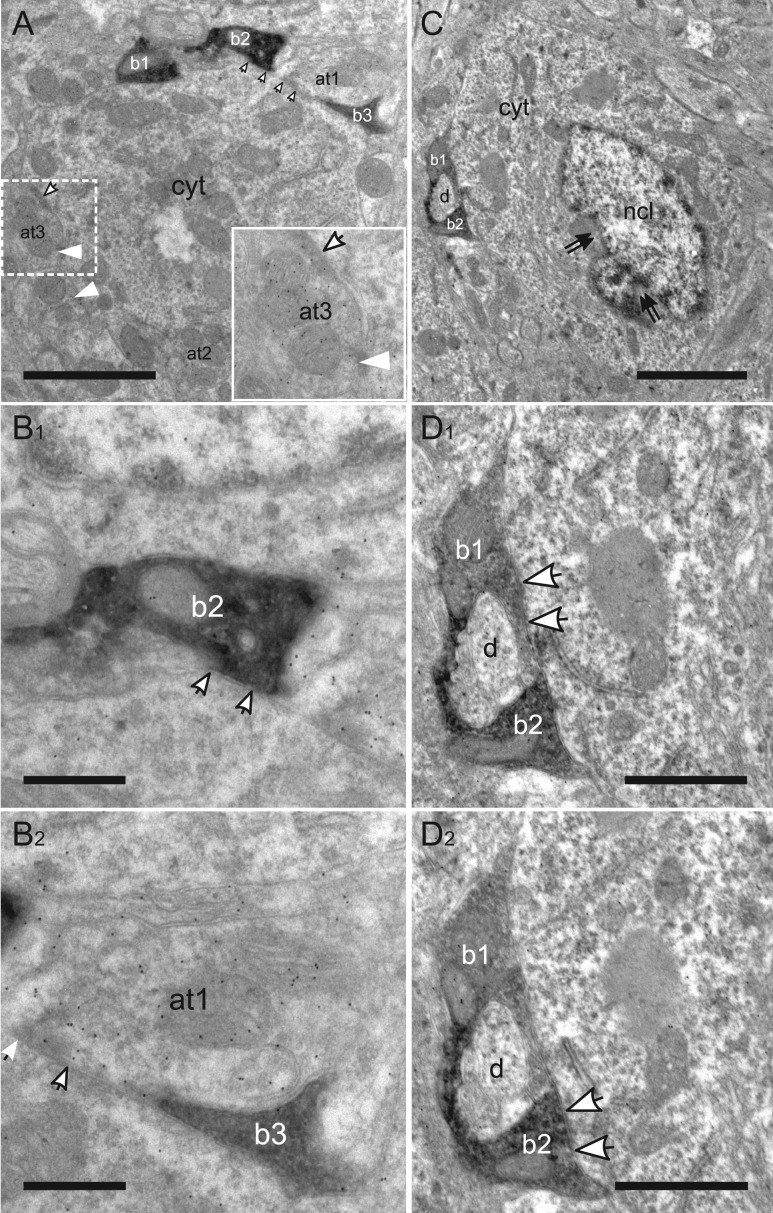
Figure 10.
Targets of VIP boutons in layer VI. (A) Low magnification image of a peripheral part of an excitatory cell soma surrounded by three VIP boutons (b1–b3) and three labeled axonal terminals (at1–at3). Puncta adherentes (white arrowheads) are differentiated from synaptic junctions (white arrows) as shown in the inset. (B1–2) Ultrastructure of somatic synapses formed by VIP bouton (b2) in (B1) and the labeled axonal terminal (at1) in (B2). (C) Low magnification image of an interneuron soma targeted by two VIP boutons (b1, b2), which enwrap a dendrite (d). Double arrows indicate indentations of the nucleus (ncl). (D1–2) Higher magnification images of two consecutive sections through the boutons in (C) showing the axosomatic synaptic junctions. cyt: cytoplasm. Scale bars = 2 μm in A; 0.5 μm in B1–2; 2.5 μm in C; 1 μm in D1–2.

