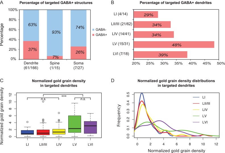
Figure 5.
GABA-labeling of the postsynaptic targets. (A) Majority of the targeted structures of VIP boutons are found to be immunonegative (GABA–). (B) Percentage of layer-specific immunopositive (GABA+) dendritic targets. Layers V and VI have the two highest immunopositive target rates. (C) Box plots of gold grain density of the VIP boutons targeting dendrites. Mann–Whitney U test shows a significant difference between superficial layers (layers I–IV) and deep layers (layers V–VI) with P value (superficial layers vs deep layers) < 0.001 and no significant difference (n.s.) within the superficial and deep layers. (D) Probability density distributions of the dataset in (C) by kernel density estimation. In superficial layers, distributions are more concentrated at around 1, which equals to background. In deep layers, however, distributions are more dispersed and most of the values are shifted to larger than 1, indicating that the labeling density in deep layers is higher than background.

