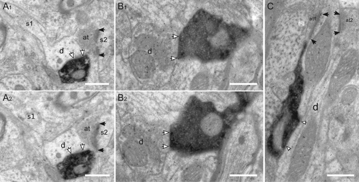
Figure 9.
Targets of VIP boutons in layer V. (A1–2) Consecutive sections show a VIP bouton which innervates an immunonegative dendrite (d; white arrows indicate the active zone). In (A1) the dendrite protrudes an elongated spine (s1). Right above there is an excitatory axospinous synapse and the postsynaptic density (black arrows in A1) has a perforation. (B) Consecutive sections show one large VIP bouton which forms a symmetric synapse with an immunopositive dendrite, densely labeled by gold grains. (C) VIP bouton targets an immunopositive dendrite which is also innervated by two excitatory axonal terminals in the vicinity (at1, at2). Scale bars = 0.5 μm in A–C.

