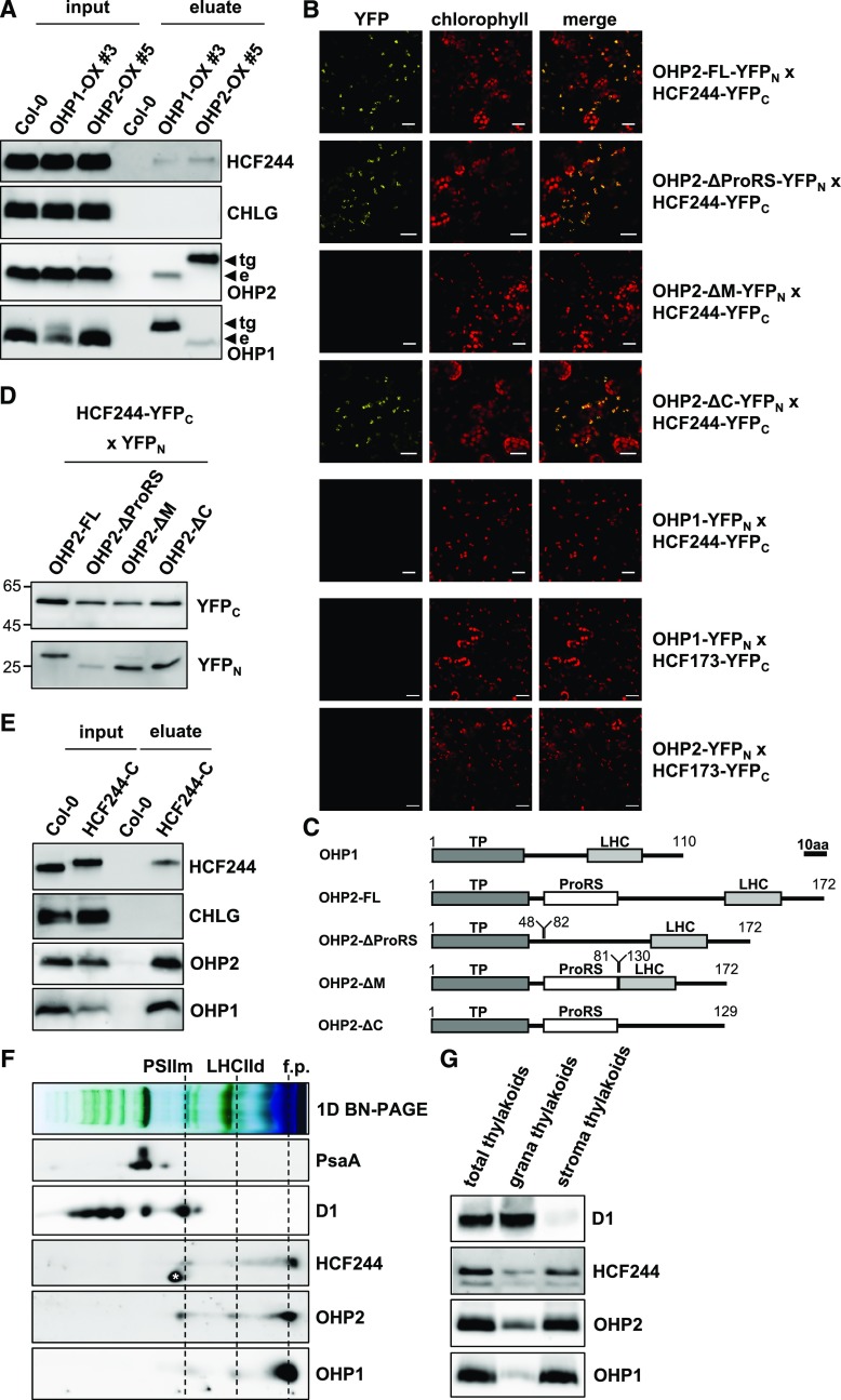
Figure 1.
OHPs interact with HCF244. A, StrepII tag pull-down analyses using OHPs as bait. Thylakoids from plants expressing OHPs fused to a C-terminal HA-strepII tag under the control of the 35S promoter (OX-lines) were solubilized with 0.5% DDM and incubated with streptavidin-coated beads. After washing and elution with 10 mm desthiobiotin, proteins were separated by Tricine-SDS-PAGE, transferred onto nitrocellulose membranes, and probed with specific antibodies. Endogenous (e) or transgenic OHP species carrying a C-terminal HA-strepII tag (tg) are indicated by arrows marking the respective signals. B, BiFC assays in N. benthamiana leaves. The indicated proteins fused to the N- and C-terminal halves of split YFP were expressed transiently in N. benthamiana leaves. Confocal microscopy on an LSM 800 confocal microscope (Zeiss) was applied to detect yellow fluorescence indicating an interaction. To confirm that the interactions occurred inside the chloroplast, chlorophyll autofluorescence also was recorded. Bars = 20 µm. C, Domain scheme of OHP species used for BiFC assays. The scheme is drawn to scale, and the scale bar represents 10 amino acids (10aa). Numbers indicate amino acids. TP, Transit peptide (predicted); LHC, transmembrane domain harboring the chlorophyll-binding motif; ProRS, Pro-rich sequence. D, Western-blot analysis of full-length (FL) and truncated OHP2 species expressed in N. benthamiana leaves for the BiFC assay. Total protein extracts were separated by SDS-PAGE, transferred onto nitrocellulose membranes, and probed with specific antibodies against the peptide tags. E, StrepII tag pull-down analyses using HCF244 as bait. Thylakoids from plants expressing HA-strepII-tagged HCF244 in the hcf244 mutant background were solubilized with 0.5% DDM, and pulldown was performed as described above. F, Analysis of complex formation of OHPs and HCF244 by BN-PAGE and subsequent second dimension SDS-PAGE. Arabidopsis wild-type thylakoids were solubilized with 1% DDM and separated on BN gels. Lanes of the BN-PAGE gels were denatured and layered on top of acrylamide gels containing 6 m urea or Tricine-SDS gels. After separation, proteins were transferred onto nitrocellulose membranes and probed with specific antibodies. PSIIm, PSII monomer; LHCIId, LHCII dimer; f.p., free proteins. The asterisk next to the HCF244 signal marks a cross reaction of the antibody. G, Subfractionation of thylakoids into grana and stroma fractions by differential centrifugation. Equal amounts of chlorophyll (4 µg) were subjected to SDS-PAGE and probed with specific antibodies.

