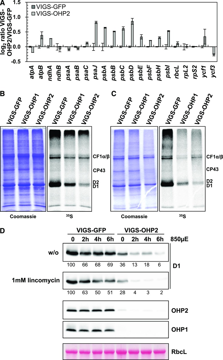
Figure 5.
Synthesis of D1 is impaired in VIGS-OHP2 plants. A, Gene expression analysis of selected plastid-encoded genes. Total RNA was reverse transcribed using random hexamer primers. SAND, ACT1, and AP2M were used as reference genes, and data were analyzed using standard curves. Data represent averages of two biological replicates, and error bars represent the sd. B and C, In vivo labeling assay of thylakoid membrane proteins. Young leaves from VIGS plants (B) or old leaves showing a pale-green phenotype described above (C) were incubated with [35S]Met in the presence of 20 µg mL−1 cycloheximide for 1 h. Crude membranes were isolated and separated by SDS-PAGE. Proteins synthesized during the incubation period were detected autoradiographically. D, In vivo protein stability of D1 in VIGS plants. Leaf discs were incubated without (w/o) or in presence of 1 mm lincomycin in high light (850 µmol photons m−2 s−1) for the times indicated. Total proteins then were separated on acrylamide gels, transferred onto nitrocellulose membranes, and probed with specific antibodies. The large subunit of Rubisco (RbcL) is shown as a loading control. Numbers below the D1 blots indicate relative amounts determined by densitometry.

