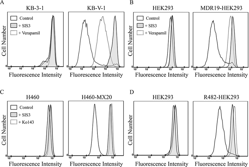
Fig. 1. SIS3 inhibits drug efflux mediated by ABCB1 and ABCG2.

The accumulation of fluorescent calcein (0.5 μM calcein-AM) in human KB-3–1 epidermal cancer cells (A, left panel) and the ABCB1-overexpressing variant KB-V-1 cancer cells (A, right panel), as well as in human HEK293 cells (B, left panel) and MDR19-HEK293, HEK293 cells transfected with human ABCB1 (B, right panel), or 1 μM of fluorescent pheophorbide A (PhA) in human H460 lung cancer cells (C, left panel) and the ABCG2-overexpressing variant H460-MX20 cancer cells (C, right panel), as well as in human HEK293 cells (D, left panel) and R482-HEK293, HEK293 cells transfected with human ABCG2 (D, right panel), was measured and analyzed immediately by flow cytometry as described previously [70]. Experiments were carried out either in the absence (solid lines) or presence of SIS3 at10 μM (shaded, solid lines) or verapamil, a reference inhibitor for ABCB1at 20 μM (A and B, dotted lines), or Ko143, a reference inhibitor for ABCG2 at 3 μM (C and D, dotted lines). Representative histograms of at least three independent experiments are shown.
