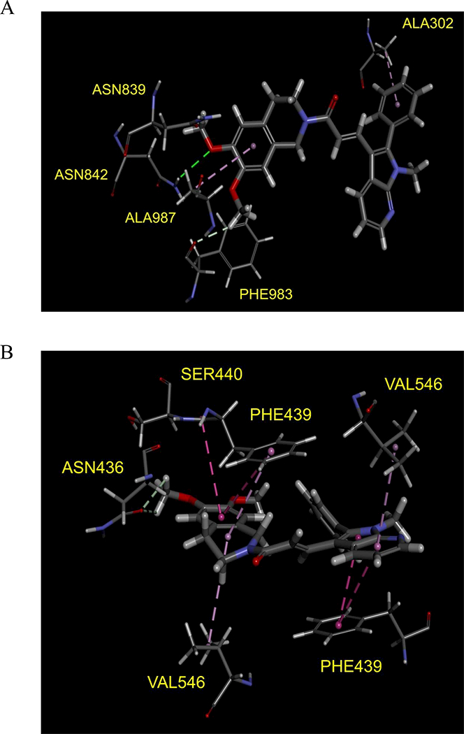
Fig. 6. Docking of SIS3 in the drug-binding pocket of ABCB1 and ABCG2.

Binding modes of SIS3 with (A) homology modeled ABCB1 and (B) ABCG2 protein structure (PDB:5NJG) were predicted by Acclerys Discovery Studio 4.0 software as described in Materials and methods. SIS3 is shown as a molecular model with the atoms colored as carbon- gray, hydrogen-light gray, nitrogen-blue and oxygen-red. The same color scheme is used for interacting amino acid residues. Dotted lines indicate proposed interactions.
