Figure 10.
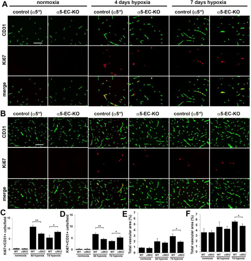
Hypoxic-induced spinal cord vascular remodeling is delayed in α5-EC-KO mice. Frozen sections of spinal cord white matter (A) or gray matter (B) taken from control or α5-EC-KO mice exposed to normoxia or 4 or 7 days hypoxia (8% O2) were examined for BEC proliferation and total vascular area, as assessed by CD31/Ki67 dual-IF. Scale bar = 100µm. C and D. Quantification of CD31+/Ki67+ proliferating BEC in the white matter (C) and gray matter (D), in which each experiment was performed with three different animals per condition, and the results expressed as the mean ± SEM of the number of CD31+/Ki67+ cells per field of view. Note that hypoxia promoted an increase in the number of proliferating BEC, though this response was delayed in α5-EC-KO mice in both white and gray matter. * p < 0.05, ** p < 0.01. E and F. Quantification of the influence of CMH on total vascular area in white matter (E) and gray matter (F). Results are expressed as the mean ± SEM in which vascular area is represented as a % of the FOV (n = 3 mice/group). Note that 7 days hypoxia promoted an increase in the total vascular area, though this response was delayed in α5-EC-KO mice both in white and gray matter. * p < 0.05.

