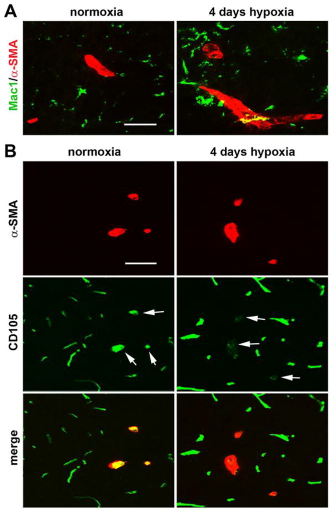
Figure 3.
Characterization of arteriogenic events in the hypoxic spinal cord. A. Dual-IF was performed on frozen sections of spinal cord gray matter taken from mice exposed to normoxia or 4 days hypoxia using antibodies specific for Mac-1 (AlexaFluor-488) and α-SMA (Cy-3). Scale bar = 25 µm. Note that in contrast to arterial vessels in the normoxic spinal cord, arteriogenic vessels in the hypoxic spinal cord were surrounded by aggregates of Mac-1-positive cells. B. Dual-IF was performed on frozen sections of spinal cord gray matter taken from mice exposed to normoxia or 4 days hypoxia using antibodies specific for CD105 (AlexaFluor-488) and α-SMA (Cy-3). Scale bar = 25 µm. Note that while α-SMA-positive arterial vessels in the normoxic spinal cord express high levels of CD105, remodeling arterioles in the hypoxic spinal cord strongly downregulate this receptor (see arrows).

