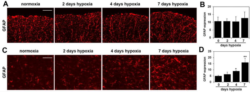
Figure 9.
The influence of chronic mild hypoxia (CMH) on astrocyte activation in the spinal cord. IF was performed on frozen sections of spinal cord white matter (A) or gray matter (C) taken from mice exposed to normoxia or 2, 4 or 7 days hypoxia using GFAP antibodies (Cy-3). Scale bar = 100 µm. B and D. Quantification of GFAP fluorescent signal at different time-points of CMH in white matter (B) and gray matter (D). Results are expressed as the mean ± SEM (n = 3 mice/group). Note that under normoxic conditions, robust GFAP expression was observed within the fibrous astrocytes of white matter, but in contrast, within the gray matter, very few GFAP-positive cells were detected. Quantification showed that CMH had no noticeable effect on the total amount of GFAP fluorescent signal in the white matter, but in the gray matter, 7 days CMH strongly increased the GFAP signal. * p < 0.05, ** p < 0.01 vs. normoxia (0 days hypoxia).

