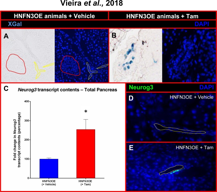
Fig 1. Analysis of the Neurog3 misexpression efficiency in HNFN3OE mice following short-term tamoxifen induction.
(A-B) β-galactosidase activity assessment in the pancreata of HNFN3OE animals treated with vehicle (A) or Tam (B) for 2 weeks. A clear activity is noted solely in ductal cells. (C) Quantitative analysis of Neurog3 transcript levels by qPCR (n = 6 animals for each condition) outlining a 2.5-fold increase in the pancreata of Tam-treated HNFN3OE animals compared to controls. Statistics were performed using the Mann-Whitney test (D-E) By means of immunohistochemical analyses using antibodies raised against Neurog3, Neurog3-expressing cells are detected within the ductal epithelium of HNFN3OE animals treated with Tam for only 2 weeks (E), whereas Neurog3+ cells cannot be detected in their vehicle-treated counterparts (D). For clarity, when required, the ductal lumen is outlined with yellow lines and islets with red lines.

