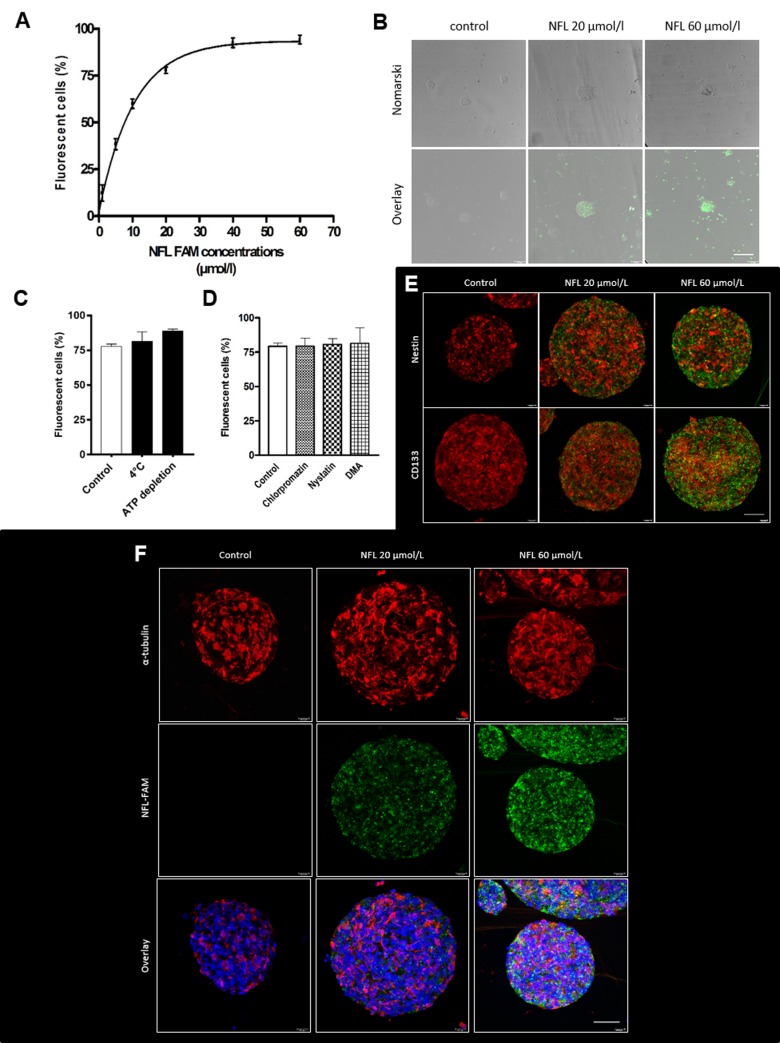
Fig 1. Uptake of the FAM-labeled NFL-TBS.40-63 peptide in hNSCs.
(A) Percentage of FAM-labeled NFL-TBS.40-63 internalized by hNSCs with increasing concentrations of peptide after 1 hour incubation at 37°C. (B) Visualization of the FAM-labeled peptide after 1 hour incubation with 20 or 60 µmol/L of peptide. Scale bar: 50 µm. Green: FAM-labeled peptide (C) Internalization of 20 µmol/L of peptide after pre-treatment at 4°C or ATP depletion or (D) in the presence of inhibitors of the endocytosis pathways. Data are presented as means ± SEM. *P<0.05. (E-F) Immunofluorescence after incubation of cells for 1 hour with 20 or 60 µmol/L FAM-labeled peptide. Cells were stained with specific neural stem cells markers: Nestin and CD133 (E) or with α-tubulin (F). Images were taken with the confocal microscope. Red: Nestin, CD133 or α-tubulin; Green: FAM-labeled peptide; Blue: DAPI. Scale bar: 50 µm.

