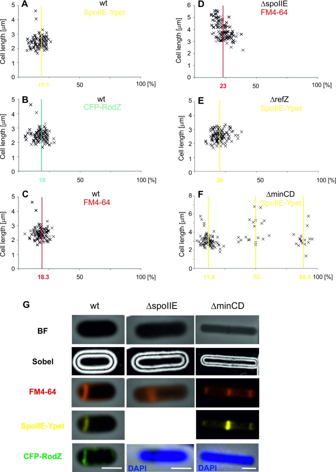
Fig 1. The sporulation septum position in wild type, ΔSpoIIE, ΔRefZ and ΔMinCD strains.
The average position of the sporulation septum is measured from the nearest cell pole, and expressed as a percentage of the total cell length (x-axis). Y-axis represents the cell length in μm. (A) The sporulation septum position in IB1538 based on the SpoIIE-Ypet signal. (B) The sporulation septum position in IB1538 based on the CFP-RodZ signal. (C) The sporulation septum position in IB1538 based on the FM4-64 membrane dye signal. (D) The sporulation septum position in the PY180 (ΔspoIIE) strain based on the FM4-64 membrane dye signal. (E) The sporulation septum position in IB1723 (ΔrefZ) based on the SpoIIE-Ypet signal. (F) The sporulation septum position in IB1724 (ΔminCD) based on the SpoIIE-Ypet signal. (G) Example images showing cell length using a Sobel filter and sporulation septum position signals from SpoIIE-Ypet, CFP-RodZ and FM4-64 in wild type, PY180 (ΔspoIIE) and IB1724 (ΔminCD) strains as described in Materials and Methods. In addition, there are DAPI staining of chromosomal DNA in PY180 (ΔspoIIE) and IB1724 (ΔminCD) strains to show that asymmetric septation started after initiation of sporulation when the nucleoid forms an axial filament from pole to pole. The scale bar represents 1 μm.

