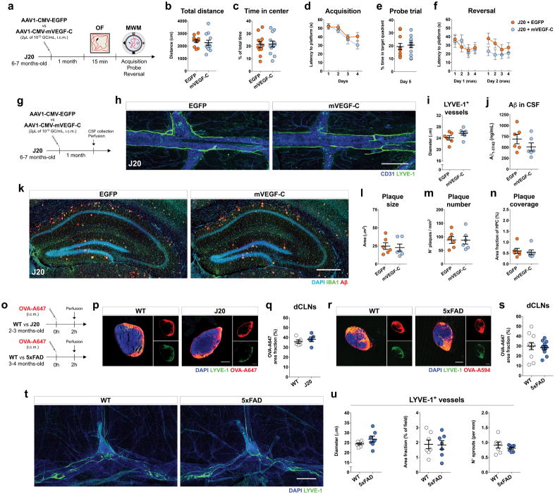
Extended Data Figure 8. Expression of mVEGF-C in the meninges of J20 mice does not ameliorate lymphatic drainage or brain amyloid pathology.
a, J20 mice were injected i.c.m. with 2 μL of AAV1-CMV-EGFP or AAV1-CMV-mVEGF-C (1013 GC/mL) at 6–7 months. One month after injection, the mice were tested in the open field (OF) and in the MWM. b, c, Total distance and % of time in the center of the OF arena was not ameliorated by treatment of J20 mice with mVEGF-C. d–f, No statistically significant differences were observed in the (d) acquisition, in the (e) probe trial or in the (f) reversal of the MWM test after 1 month of mVEGF-C. Data in b–f is presented as mean ± s.e.m., n = 11 in EGFP, n = 12 in mVEGF-C; two-tailed Mann-Whitney test was used in b, c and e and repeated measures two-way ANOVA with Bonferroni’s post-hoc test was used in d and f; data results from a single experiment. g, J20 mice were treated with EGFP or mVEGF-C and, 1 month later, CSF, meninges and brain were collected for analysis. h, Representative images of DAPI (blue) and LYVE-1+ lymphatic vessels (green) in the superior sagittal sinus of mice treated with either EGFP or mVEGF-C (scale bar, 500 μm). i, AAV1-mediated expression of mVEGF-C did not affect meningeal lymphatic vessel diameter. j, Levels of Aβ in the CSF measured by ELISA remained unaltered after mVEGF-C treatment. k, Representative images of dorsal hippocampus (scale bar, 500 μm) of J20 mice of EGFP or mVEGF-C groups stained with DAPI (cyan) and for IBA1 (green) and Aβ (red). l–n, No changes were observed in amyloid plaque (l) size, (m) number or (n) coverage between the groups. Data in i, j and l–n is presented as mean ± s.e.m., n = 6 per group; two-tailed Mann-Whitney test was used in i, j and l–n; data in g–n results from a single experiment. o, J20 mice (2–3 months-old) and 5xFAD mice (3–4 months-old), and respective age-matched WT littermate controls, were injected with fluorescent OVA-A647 (i.c.m.) in order to measure drainage into the dCLNs. p, Representative images of DAPI (blue) and LYVE-1 (green) staining in dCLNs of WT and J20 mice (scale bar, 200 μm) 2 h after injection of OVA-A647 (red). q, Quantification of OVA-A647 area fraction (%) in the dCLNs shows equal levels of tracer in mice from both genotypes (mean ± s.e.m., n = 5 per group; two-tailed Mann-Whitney test; representative of 2 independent experiments). r, Representative images of DAPI (blue) and LYVE-1 (green) staining in dCLNs of WT and 5xFAD mice (scale bar, 200 μm) 2 h after injection of OVA-A594 (red). s, Quantification of OVA-A594 area fraction (%) in the dCLNs shows equal levels of tracer in mice from both genotypes (mean ± s.e.m., n = 11 per group; two-tailed Mann-Whitney test; data was pooled from 2 independent experiments). t, Representative images of DAPI (blue) and LYVE-1 (green) staining in meningeal whole-mounts of WT and 5xFAD mice at 3–4 months (scale bar, 1 mm). u, Measurement of LYVE-1+ vessel diameter, area fraction and number of sprouts (per mm of vessel) showed no differences between genotypes (mean ± s.e.m., n = 7 per group; two-tailed Mann-Whitney test; data was pooled from 2 independent experiments).

