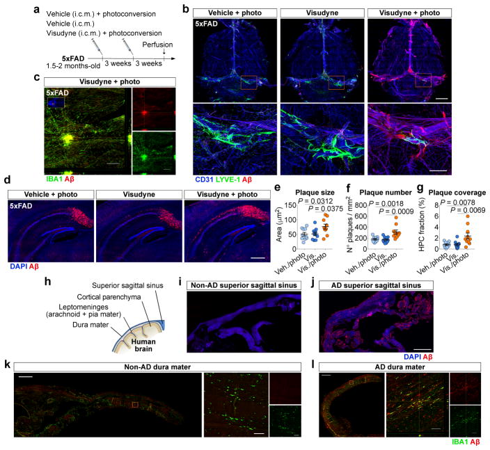
Figure 3. Ablation of meningeal lymphatics aggravates amyloid pathology in AD transgenic mice.
a, Young-adult 5xFAD mice were submitted to meningeal lymphatic ablation or control procedures. Procedures were repeated 3 weeks later and amyloid pathology was assessed 6 weeks after initial treatment. b, Staining for CD31/LYVE-1/Aβ in meninges (scale bar, 2 mm; inset scale bar, 500 μm). c, Orthogonal view of IBA+ macrophages clustering around an amyloid plaque in meninges of a 5xFAD with ablated lymphatics (scale bar, 200 μm). d, Representative images of DAPI/Aβ in the hippocampus of 5xFAD mice from each group (scale bar, 500 μm). e–g, Quantification of amyloid plaque (e) size, (f) number and (g) coverage in the hippocampus of 5xFAD mice. Data in e–g is presented as mean ± s.e.m., n = 10 per group; one-way ANOVA with Bonferroni’s post-hoc test was used in e–g; a–g is representative of 2 independent experiments. h, Staining for amyloid pathology was performed in human non-AD and AD brains (Extended data Fig. 9) and different meningeal layers. i, j, Meningeal superior sagittal sinus tissue of (i) non-AD or (j) AD patients stained with DAPI/Aβ (scale bar, 2 mm). k, l, Meningeal dura mater tissue of (k) non-AD or (l) AD patients, stained for IBA1/Aβ (scale bars, 1 mm; orthogonal view inset scale bars, 50 μm). Data in h–l results of n = 8 non-AD samples and n = 9 AD samples and is representative of 2 independent experiments.

