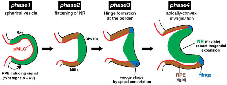
Figure 5.

Alternative hypothesis for optic cup formation. Phase 1: Apical contraction (red) produces spherical optic vesicle. pMLC = phosphorylated myosin light chain. Phase 2: Relaxation in central region produces retinal placode (green). Phase 3: Apical contraction at placode border causes sharp bending (blue) and slight invagination. Phase 4: Growth of placode region deepens invagination. Republished with permission from Eiraku et al. (2012).
