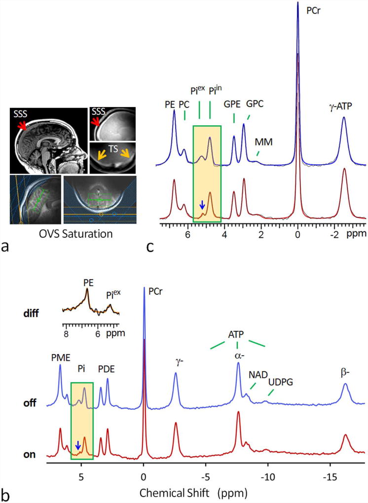
FIG. 1.
(a) 7T MR head images showing the location of superior sagittal sinus (SSS), transverse sinus (TS), and the 31P saturation slabs for outer-volume-suppression (OVS). The green box in the images represents the 1H shimming area. (b) Fully-relaxed 7T 31P MR spectra at long TR of 30 sec, acquired from resting human brain using a pulse-acquire sequence with (red trace) and without (blue trace) OVS saturation. Note the marked attenuation of the signals at Piex and PE upon OVS saturation, while other metabolite 31P signals remain unchanged in reference to PCr at 0 ppm. Also note the asymmetric lineshape of the attenuated peak at PE (inset) as featured in typical membrane phospholipid signal due to chemical shift anisotropy effect. (c) The fitted 31P MR spectra in the chemical shift region between -4.0 and 8.0 ppm (red trace: OVS on; blue trace: OVS off). Abbreviation: PE, phosphoethenolamine; PC, phosphocholine, GPE, glycerophosphoethanolamine; GPC, glycerophosphocholine; Piin and Piex, intra- and extracellular inorganic phosphate; PCr, phosphocreatine; ATP, adenosine triphosphate; NAD, nicotinamide adenine dinucleotide; UDPG, uridine diphosphate glucose; PME, phosphomonoester; PDE, phosphodiester; MM, macromolecules (likely from mobile membrane phospholipids).

