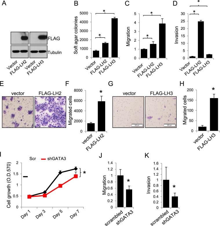
Figure 3.
Expression of LH2 or LH3 promotes lung cancer cell growth and migration. (A–D) Western blotting (A), soft agar colony formation, (B), migration (C), and invasion (D) assays for 393 P cells stably expressing FLAG-tagged LH2 and LH3 or empty vectors. (E) Microscopic images for the GATA3-depleted 344SQ cells that stably express vector or FLAG-tagged LH2 and migrated through non-coated transwells. (F) The number of migrated cells were counted and expressed as mean + stdv. (G) Microscopic images for the HCC827 cells that stably express vector or FLAG-tagged LH3 and migrated through non-coated transwells. (H) The number of migrated cells were counted and expressed as mean + stdv. (I–K) MTT (I), migration (J), and invasion (K) assay for 393 P cells transfected with scrambled (scr) or GATA3 shRNAs. Note: in all figures, *indicates t-test p < 0.05.

