Fig. 2.

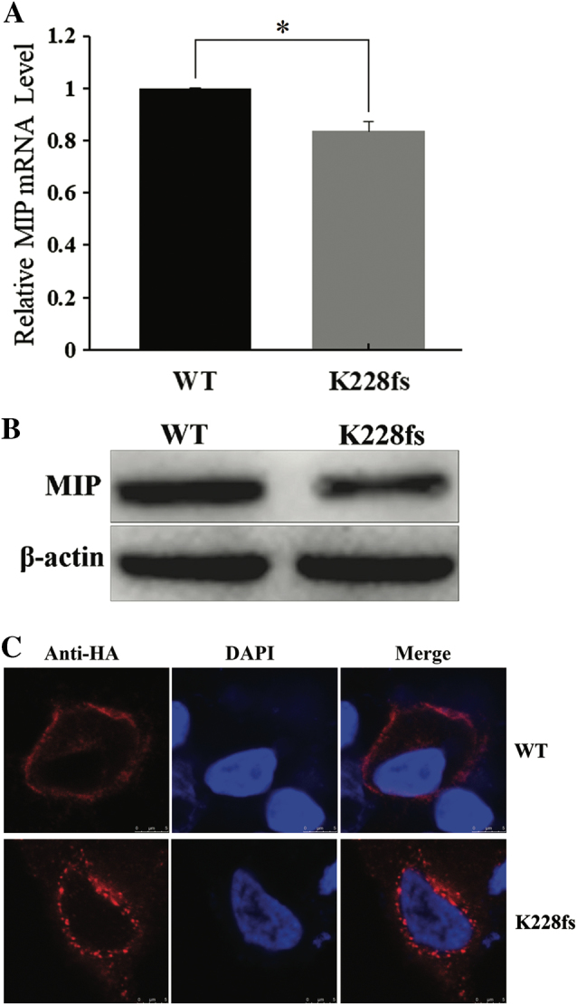
Expression of WT-MIP and K228fs-MIP in cultured HeLa cells. a The mRNA transcription level of K228fs-MIP was decreased compared with that of WT-MIP according to quantitative real-time PCR (T-TEST, two-sided, *P = 0.0052, error bars represent standard deviation, SD). All of the samples were analyzed in four replicates and normalized to median β-actin expression. b Western blots were performed as indicated and showed that the protein expression of K228fs-MIP was lower than that of WT-MIP. The expression of β-actin was used as the control. c Subcellular localization of HA-WT-MIP and HA-K228fs-MIP were determined after transient transfection in cultured HeLa cells. Confocal laser scanning images showed the distribution of HA-tagged MIP (red) and DAPI-stained nuclei (blue). HA-WT-MIP was detected mainly in the plasma membrane with less observed in the cytoplasm, but K228fs-MIP was largely localized in the cytoplasm. Scale bar: 5 μm
