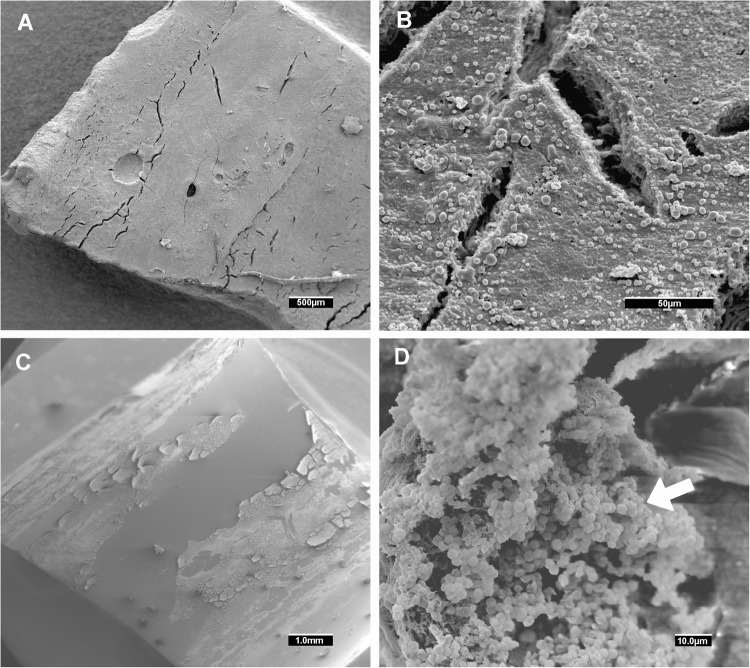
Figure 4.
Representative scanning electron microscopy of in vivo MRSA biofilm (A) Isolate 1 showing an in vivo detached biofilm at low magnification. Sometimes the sample processing for scanning electron microscopy released the biofilm cluster from the endotracheal tube surface. At higher magnification (B), cocci morphologies can be distinguished. The pig from which we obtained Isolate 1 was treated with vancomycin. (C) Isolate 45 (from a placebo treated pig) showing an in vivo biofilm attached to the endotracheal tube at low magnification. (D) at higher magnification, a cocci biofilm cluster was found (white arrow). Abbreviations: MRSA, methicillin-resistant Staphylococcus aureus.

