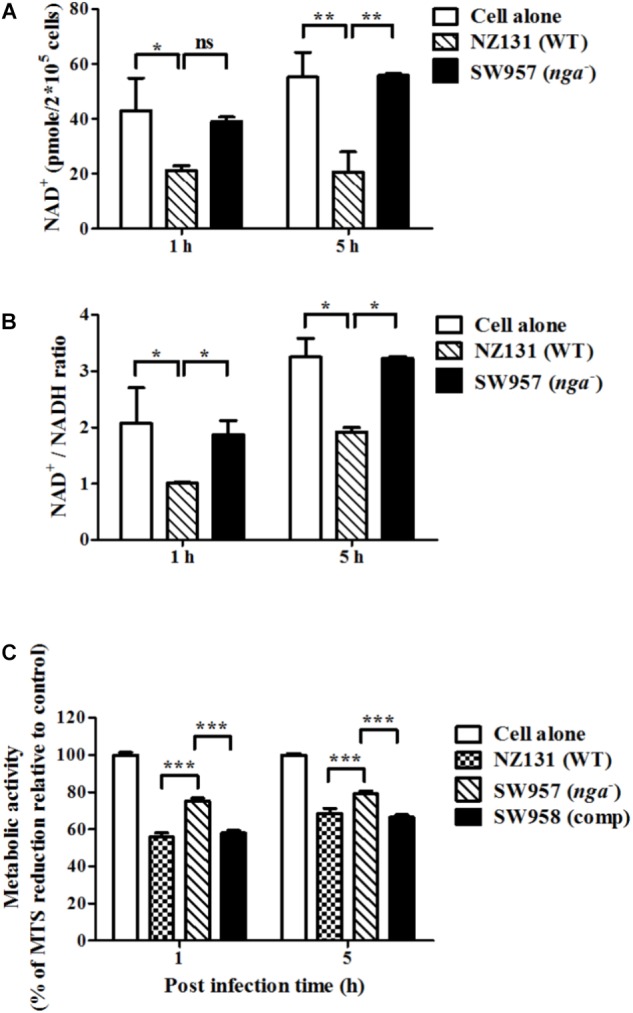
FIGURE 3.

NADase depletes intracellular NAD+ content within infected endothelial cells. (A) The intracellular NAD+ content in endothelial cells after infection with NZ131 or the nga mutant. Intracellular NAD+ was extracted from GAS-infected HMEC-1 cells during infection and the content was analyzed by a NAD+/NADH quantification kit. (B) The NAD+/NADH ratio in endothelial cells after infection with NZ131 or the nga mutant. The NAD+/NADH ratio of cell lysates was calculated following a formula: [(NADtotal – NADH)/NADH]. (C) The cell viability of infected cells was analyzed by metabolic activity of mitochondria using the MTS assay. The data represent the means ± SEM of at least three independent experiments. ∗p < 0.05; ∗∗p < 0.01; ∗∗∗p < 0.001 (two-way ANOVA).
