
Fig. 1.
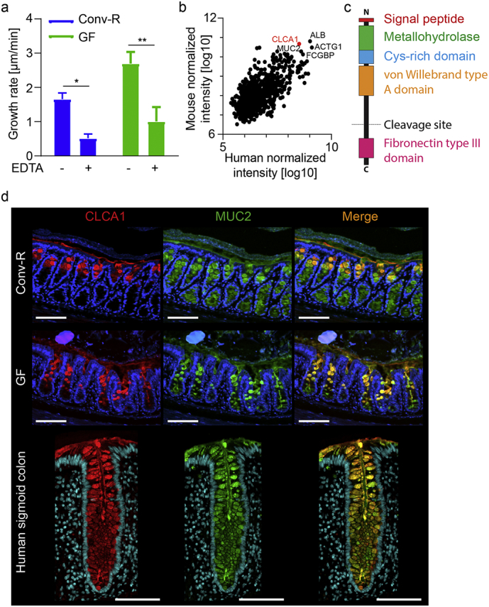
CLCA1 is a candidate to be the metalloprotease involved in mucus growth in WT and GF colon. (a) Distal colon mucus growth rate measured in Conv-R and GF mice without (−) and with (+) 10 mM EDTA applied apically (n = 4–6). (b) Proteome analysis of mouse and human colonic mucus. The five shared most abundant proteins are labeled with their gene names. CLCA1 is marked in red (mouse n = 7, human n = 47). (c) Schematic representation of CLCA1 with marked domains. (d) Immunostainings of CLCA1 (red) and MUC2 (green) in Conv-R and GF mice, and in human sigmoid colon sections. DNA was stained with Hoechst (blue) (representative of 9–15). Scale bar is 100 μm. Data is presented as mean ± SEM; * p ≤ .05, ** p ≤ .01, with two-way ANOVA with Sidak's multiple comparisons test (MCT).