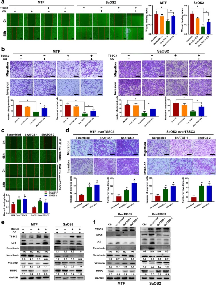
Fig. 5.
TSSC3 inhibits osteosarcoma cell migration and invasion associated with autophagy. a A wound healing assay was conducted for over-Ctrl and TSSC3-overexpression MTF or SaOS2 cells treated with or without CQ (8 μM, 12 h) for 48 h. * P < 0.05, Scale bars: 200 μm. b Transwell migration and invasion assays indicate blockage of the autophagy flux by CQ (8 μM) alleviates TSSC3-induced inhibition of osteosarcoma cells migration and invasion in vitro. * P < 0.05, Scale bars: 200 μm. c ATG5 knockdown blocked TSSC3-induced suppression of cell migration. MTF-TSSC3-Scrambled/shATG5–1 or 2 and SaOS2-TSSC3-Scrambled/shATG5–1 or 2 cells migration abilities were measured using a wound healing assay for 48 h. * P < 0.05, compared with the MTF-Scrambled group, # P < 0.05, compared with the SaOS2-Scrambled group, Scale bars: 200 μm. d Transwell migration and invasion assays with cells treated as mentioned above also show that ATG5 knockdown blocked TSSC3-induced suppression of cell migration. * P < 0.05, Scale bars: 200 μm. e-f Representative images of western blotting analysis of autophagy-related proteins and epithelial-mesenchymal transition and invasion-related proteins levels in MTF and SaOS2 cells. Cells were treated with or without TSSC3-overexpression and TSSC3-induced autophagy was blocked using CQ or shATG5. Protein levels on the western blots were quantified by densitometry of E-cadherin, N-cadherin, Vimentin and MMP2, normalized to GAPDH and given as the fold change compared with that in the control groups. NG: Not given. Quantification data was in Additional file 3: Figure S5

