Fig. 6.
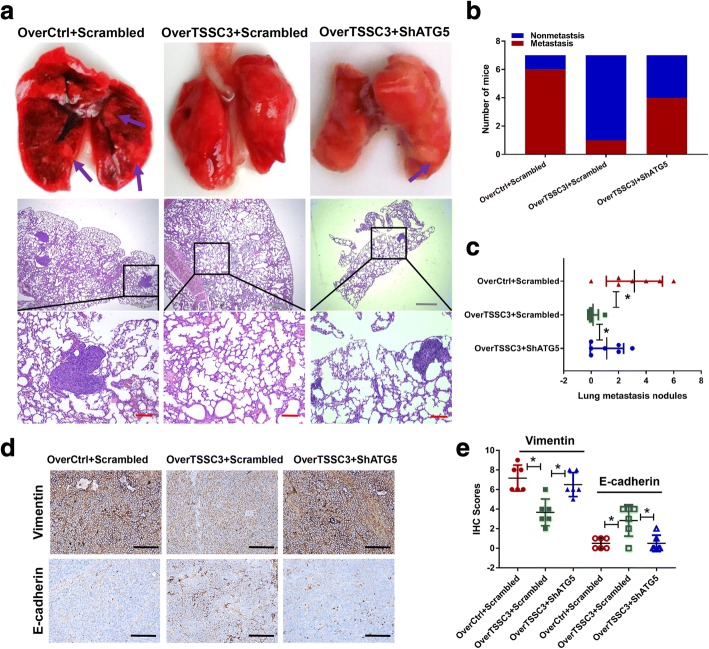
Autophagy contributes to TSSC3-induced lung metastasis suppression in vivo. MTF cells were transfected as indicated and injected into the tail vein of nude mice to establish an in vivo model of lung metastasis. a Representative macroscopic and microscopic images (H&E staining) of the lungs. Purple arrows indicate possible metastatic lesions. Gray scale bars: 500 μm, red scale bars: 100 μm. b Percentage of mice bearing lung metastases in each group (n = 7). c Numbers of lung metastatic nodules quantified on the section with H&E staining of the lungs. *P < 0.05. d-e E-cadherin and Vimentin protein expression in xenograft tumors, quantified by immunohistochemistry (IHC). The quantification of the IHC scores is shown in e. *P < 0.05

