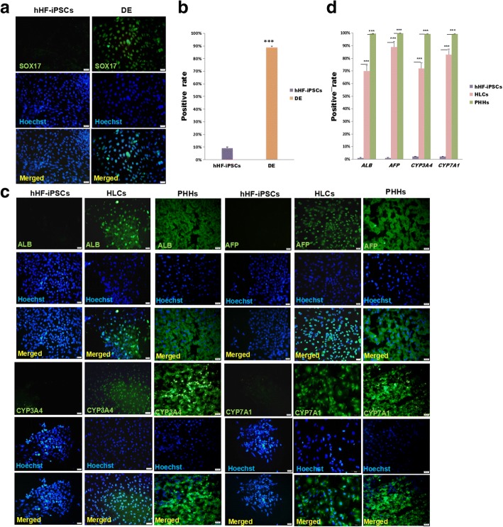
Fig. 2.
Protein levels in DE, HLCs, and PHHs. a Protein levels in DE. hHF-iPSCs and DE immunostained for SOX17 (green) and counterstained with DAPI (blue). b Quantification of SOX17 expression in hHF-iPSCs and DE (***P < 0.001; n ≥ 3). c Protein levels in HLCs. hHF-iPSCs, HLCs, and PHHs immunostained for ALB, AFP, CYP3A4, and CYP7A1 (green), and counterstained with DAPI (blue) d Quantification of ALB, AFP, CYP3A4, and CYP7A1 expression in hHF-iPSCs, HLCs, and PHHs (***P < 0.001; n ≥ 3). Scale bars = 20 μm. AFP alpha-fetoprotein, ALB albumin, CYP cytochrome P450, DE definitive endoderm, hHF-iPSC human hair follicle-derived induced pluripotent stem cell, HLC hepatocyte-like cell, PHH primary human hepatocyte

