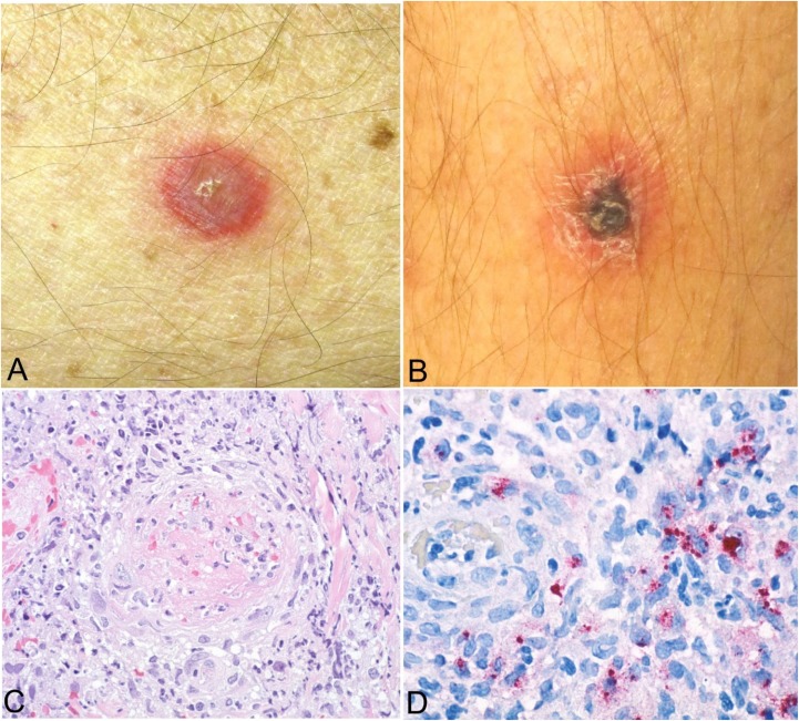
Figure 3.
Clinical and histopathological appearance of primary lesions of African tick bite fever (ATBF). Lesions occur at the tick-bite site and begin as an erythematous plaque or vesicle (A) that enlarges and develops into an inoculation eschar, characterized by a central, 0.5–3.0-cm ulcer covered by a brown-black crust and typically surrounded by an annular red halo (B). Eschars of ATBF occur most commonly on the lower extremities and frequently as multiple lesions.10 The histological appearance of eschars includes dermal and epidermal necrosis with predominantly lymphohistocytic perivascular inflammatory cell infiltrates in the superficial and deep dermis, often associated with thrombosis and mural necrosis of inflamed small vessels (hematoxylin and eosin stain, original magnification ×100) (C). Immunohistochemical staining typically reveals abundant rickettsial antigens (red) in the infiltrates that surround the vessels (immunoalkaline phosphatase with naphthol fast-red and hematoxylin counterstain, original magnification ×158) (D). Images (A) and (B) courtesy of Charles Thurston, MD, and Lester Libow, MD; Images (C) and (D) courtesy of Sherif Zaki, MD, PhD.

