FIGURE 1:
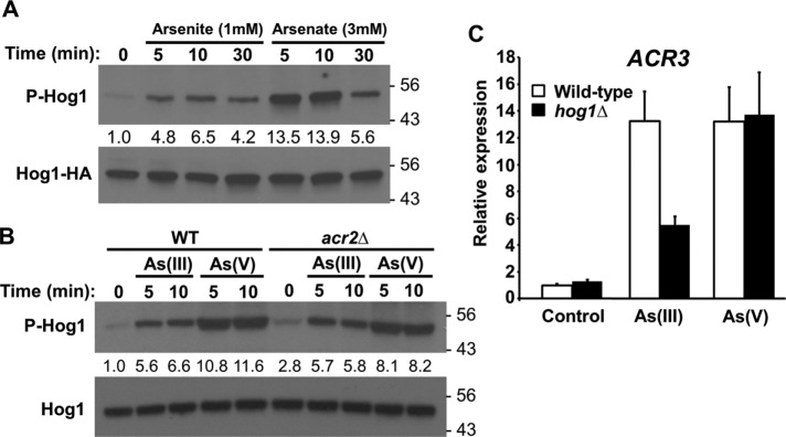
As(III) and As(V) activate Hog1. (A) Activation of Hog1 by As(III) and As(V). Wild-type yeast cells (DL3187) expressing Hog1-HA (from p3225) were treated as indicated and extracts were separated by SDS–PAGE prior to examination by immunoblot for activated (phospho)-Hog1 and total Hog1-HA. Fold activation values are relative to the untreated sample and normalized to the Hog1-HA input. Molecular mass markers (in kDa) are on the right. An α-Hog1 antibody was used for all subsequent immunoblots to detect endogenous Hog1. (B) Activation of Hog1 by As(V) does not require its conversion to As(III). Wild-type or acr2Δ cells (DL4341) were treated as indicated with 1 mM As(III) or 3 mM As(V) and examined for activation of endogenous Hog1. Fold activation values are relative to the untreated wild-type sample and normalized to the Hog1 input. Note that the acr2Δ mutant displays slightly higher basal Hog1 phosphorylation than the wild type. (C) Induction of the arsenic protection gene ACR3 by As(III), but not by As(V), is partially dependent upon HOG1. ACR3 expression was measured by quantitative real-time PCR in wild-type cells and hog1Δ cells (DL3158) after treatment with 1 mM As(III) or 3 mM As(V) for 5 min. Each data point represents the mean and SD of three biological replicates.

