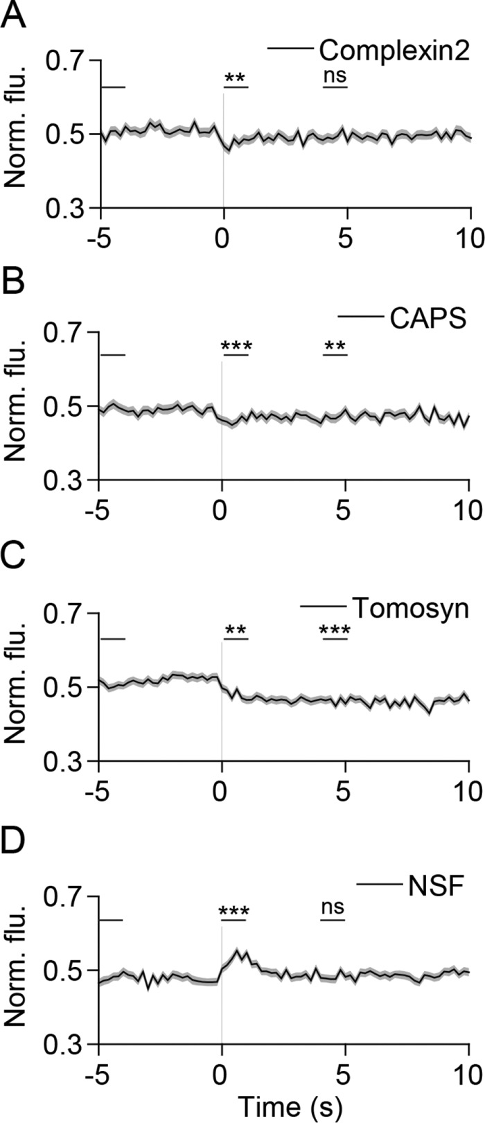
FIGURE 4:

SNARE modulators exhibit diverse behaviors during SLMV fusion. (A–C) Average time-lapse traces of normalized fluorescence intensities for (A) complexin2-mCherry (202 events, seven cells), (B) CAPS-mKate2 (232 events, five cells), (C) mCherry-tomosyn (289 events, nine cells), and (D) NSF-mCherry (274 events, five cells). Individual event traces were time aligned to 0 s (vertical black line), which corresponds to the fusion frame in the green channel. Standard errors are plotted as shaded areas around the average traces. **p ≤ 0.01, ***p ≤ 0.0001, ns, not significant, when compared with baseline.
