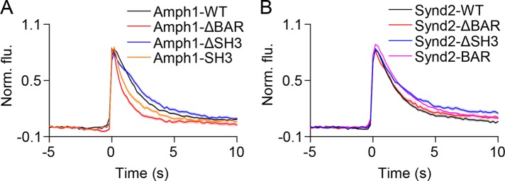
FIGURE 7:
Amphiphysin1 and syndapin2 mutants slow the loss of VAChT-pH from fusion sites. (A) Average time-lapse traces of normalized VAChT-pH fluorescence intensities in PC12 cells coexpressing VAChT-pH and (A) WT or mutant amphiphysin1 and (B) WT or mutant syndapin2 constructs. Individual event traces were time aligned to 0 s, which corresponds to the frame of fusion. Standard errors are plotted as shaded areas around the average traces.

