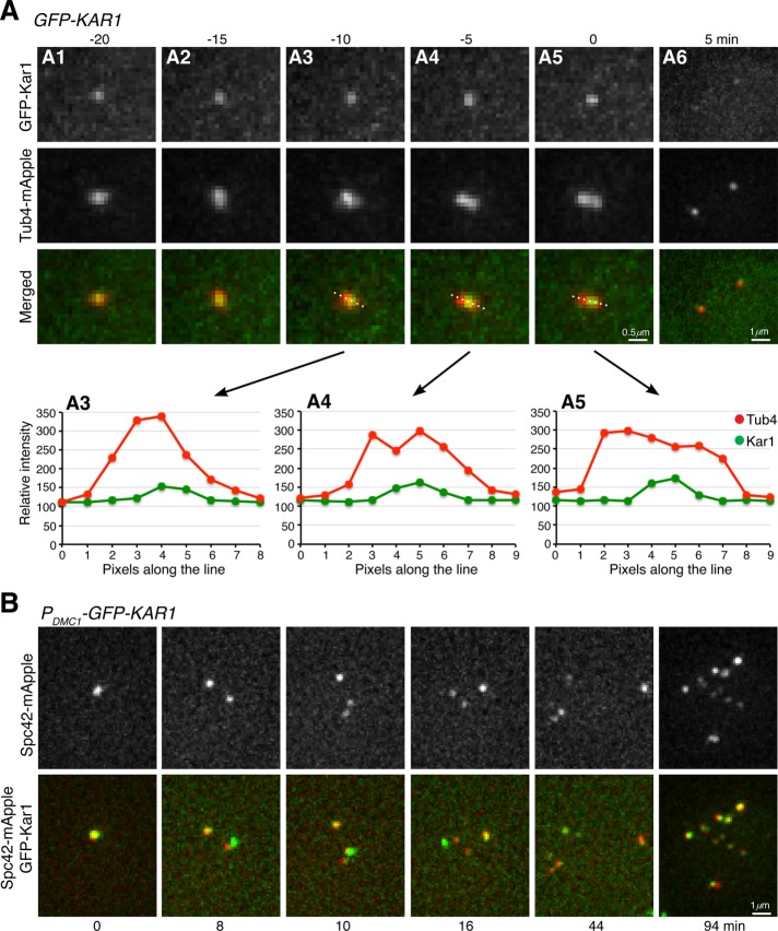
FIGURE 2:
Localization of Kar1 to the half-bridge area during yeast meiosis. (A) Time-lapse live-cell microscopy was performed as in Figure 1B. Single optical sections are shown. Graphs of line-scan pixel intensity shown in A3 to A4 correspond to the top images. Time zero refers to the point of SPB separation in meiosis I. Note that GFP-Kar1 localizes to the central region of two separating SPBs, which are marked by Tub4-mApple. (B) Colocalization of Spc42 with Kar1 in yeast meiosis. Time-lapse live-cell microscopy was performed as in A. Time interval was set at 2 min; projected images from 12 optical sections are shown. Time zero refers to the point of the first round of Spc42 separation in meiosis I. Note that all Spc42-mApple foci colocalize with GFP-Kar1.

