FIGURE 7:
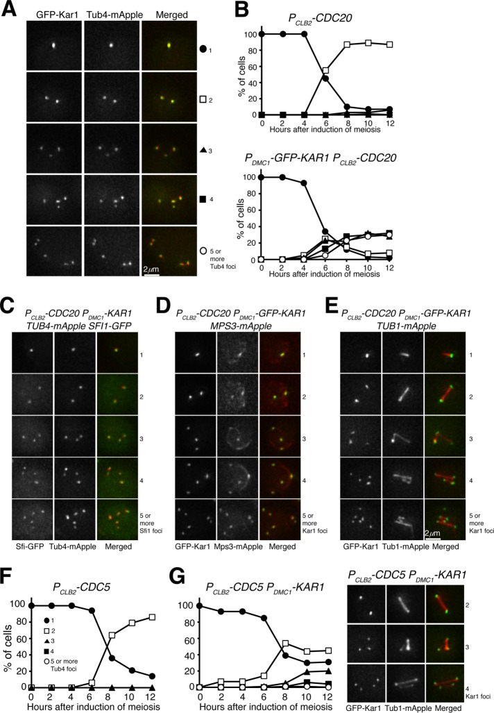
The execution point of Kar1-mediated SPB duplication in yeast meiosis. (A) Representative images showing GFP-Kar1 and Tub4 localization in PDMC1-GFP-KAR1 PCLB2-CDC20 cells. Depletion of Cdc20 arrests yeast cells at metaphase I. (B) Quantification of SPB separation in PCLB2-CDC20 and PDMC1-GFP-KAR1 PCLB2-CDC20 cells. Note the formation of supernumerary SPBs in PDMC1-GFP-KAR1 PCLB2-CDC20 cells. (C) Representative images showing Sfi1 localization in PDMC1-GFP-KAR1 cells at metaphase I. Note that Sfi1-GFP colocalizes with Tub4-mApple. (D) Representative images showing Mps3 localization in PDMC1-GFP-KAR1 cells at metaphase I. (E) Representative images showing Tub1 localization in PDMC1-GFP-KAR1 cells at metaphase I. Projected images are shown (C–E). (F) Quantification of SPB separation in Cdc5-depleted (PCLB2-CDC5) cells in meiosis. (G) Quantification of SPB separation in PDMC1-GFP-KAR1 PCLB2-CDC5 cells. Representative images of GFP-Kar1 and Tub1-mApple are shown to the right. The time-course experiments were repeated, and data from one representative experiment are shown.

