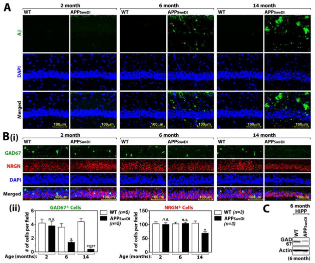
Fig. 1. Age dependent Aβ accumulation and degeneration of GABAergic neurons in hippocampus.
A. Aβ42 accumulation in CA1 of Hippocampus of WT and APPSwDI mice (2, 6, and 14 months old) were analyzed by immunofluorescent staining with BC05 antibody. DAPI was used for staining for nucleus. B. Age dependent loss of GABAergic neurons (GAD67+ cells) or pyramidal neurons (neurogranin/NRGN) were investigated (i). The graph shows the # of GABAergic or pyramidal neurons (ii). C. The expression of GAD67 in hippocampus (HIPP) was analyzed by Western blot. All columns are means of individual data and T-bars are standard error mean: n.s. P > 0.05. *. P ≤ 0.05. ****. P ≤ 0.0001 as compared to WT mice.

