
Figure 2. Genetic suppression of TAZ inhibits aRMS cell growth, decreases cell proliferation, and increases apoptosis.
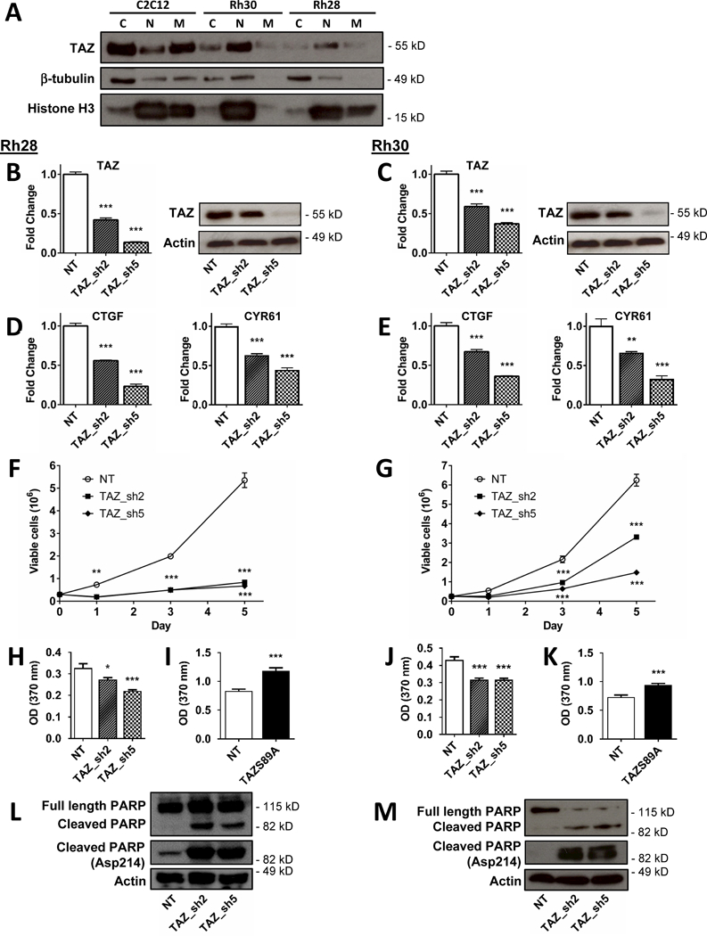
(A) Cell fractionation reveals enrichment of nuclear (active) TAZ in Rh28 and Rh30 aRMS cells, compared to primarily cytoplasmic/membrane expression in C2C12 murine myoblasts. β-tubulin and histone H3 are used as markers of cytoplasmic and nuclear expression. (B, C) Lentiviral-mediated suppression of TAZ in Rh28 and Rh30 cells shows consistent knockdown as measured by qRT-PCR and immunoblot. TAZ knockdown also leads to (D, E) suppression of TAZ target genes CTGF and CYR61 as measured by qRT-PCR, (F, G) decreased cell growth as measured by cell counting in culture, and (H, J) decreased proliferation as measured by BrdU incorporation. Conversely, aRMS cells expressing constitutively active TAZ (TAZS89A) show increased proliferation (I, K). TAZ suppression also led to increased apoptosis, as measured by immunoblots of both full length and cleaved PARP (L, M). Actin used as loading control. C, cytoplasmic; N, nuclear; M, membrane; NT, non-targeting scrambled control vector. *, P<0.05; **, P<0.01; and ***, P<0.001.