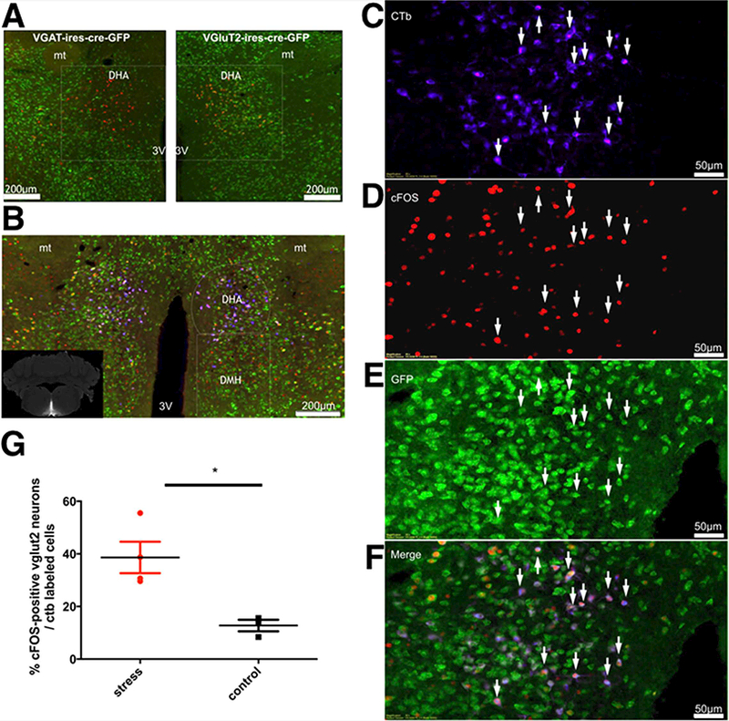
Figure 1. Identification of RPa projecting-DHA neurons activated during stress.

(A) Neurons in the dorsal hypothalamic area (DHA) were retrogradely labeled (red) by an injection of Cholera Toxin subunit b (CTb) in the medullary raphe pallidus (RPa) of Vgat-IRES-cre-GFP mice, n=4 and Vglut2-IRES-cre-GFP mice, n=7. Note that the retrograde label is highly colocalized with Vglut2 (yellow color in right panel). (B) Many of the Vglut2 neurons (green) that project to the RPa (blue; see inset for injection site) also contain c-fos (red). These neurons appear purple in this triple color scheme. Panels C-F show the left side of panel D at higher magnification. (G) Approximately 39 ± 6 % of the neurons retrogradely labeled from the RPa also containd cFOS and VGLUT2 in animals subjected to cage exchange stress, but only about 13 ± 2% in non-stressed control animals. *Statistical difference between groups (stress, n=4 and control, n=3) (p=0.0166, t-test=3.538 df=5). Abbreviations: 3V, third ventricle; mt, mammillothalamic tract.
