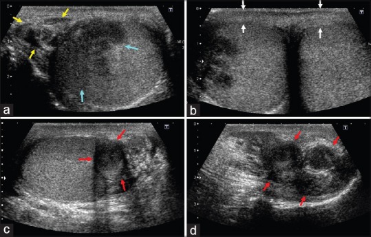
Figure 2.

(a) Transverse gray-scale image reveals enlargement and heterogeneous echo texture of tail of the right epididymis (yellow arrows) on right with direct extension to the testicle (blue arrows). (b) Thickening of scrotal skin (white arrows). (c and d) Uninvolved left testicle is with enlarged and nodular left epididymis (red arrows in c and d, longitudinal scan), suggestive of tuberculous epididymo-orchitis
