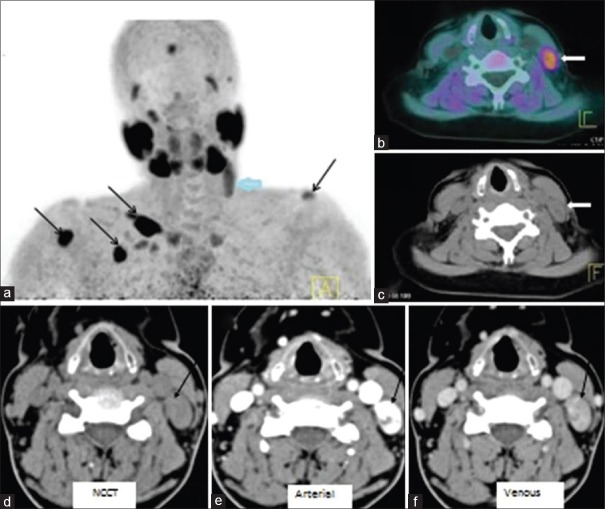
Figure 4.
FCH PET/CT (a) MIP shows focal uptake in left upper neck (thick arrow). Also, multiple foci of increased tracer uptake are seen over left shoulder, chest wall, and right arm regions (black arrows) – brown tumours. Fused PET/CT (b) and CT (c) images show soft tissue nodule in the left lateral neck, posterior to left sternocleidomastoid muscle (thick arrow). 4D-CT (d–f) shows an intensely enhancing lesion (arrow) on arterial phase with washout in venous phase posterior to the left jugular vein at thyroid cartilage level suggesting ectopically located parathyroid adenoma

