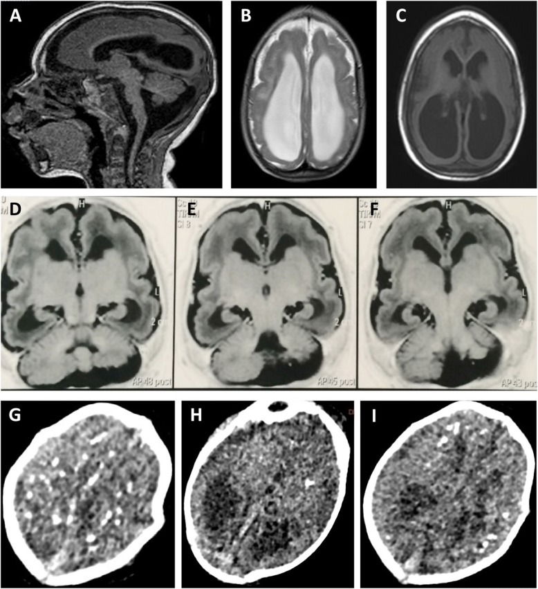
Fig. 1.
Brain Imaging after birth. a MRI Sagittal T1 weighted image: hypogenesis of the corpus callosum, enlarged cisterna magna, and ventriculomegaly. b MRI Axial T2 weighted image: simplified frontal gyral pattern, ventriculomegaly. c Axial T1 weighted image: pachygyria/lissencephaly in the frontal lobe, ventriculomegaly. d, e, f MRI Axial T1 weighted image: simplified frontal gyral pattern, ventriculomegaly, cerebellum hemisphere hypoplasia. g, h, i Axial non-contrast CT image: multiple bilateral calcifications in the junction between cortical and subcortical white matter, ventricular enlargement

