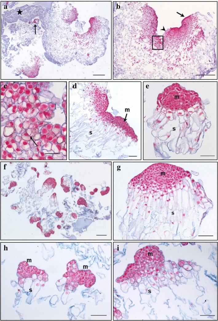
Fig. 4.
Histology of embryonal masses from primary and secondary lines of the genotype SD4. a, b, c / SD4; d, e / SD4–2; f, g / SD4–6; h, i / SD4–8. a, b – structures resembling the polyembryogenic centers (PECs) Arrow in a points to small somatic embryos (SEs) and the star marks the dead material in the end of suspensor region; arrow in b points to the smooth surface of protoderm and arrowhead marks the place where protoderm is missing; the detail of the framed region in b is shown in c; arrow in c points to the brown cells with phenolic content located in the meristem-like region; d – PEC; e – singulated SEs, f – cluster of small SEs and PECs; g – well-organized SEs; h – small SEs; i – small PECs; m – meristem of PECs or singulated SEs, s – suspensor. Paraffin sections stained with Alcian Blue/Nuclear Fast Red Scale bars: a, b = 500 μm; c = 50 μm; d, e, g, h, I = 100 μm; f = 200 μm

