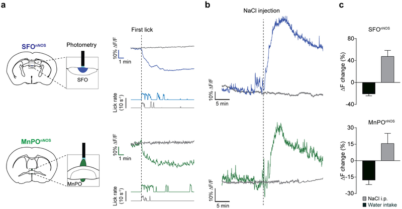
Extended Data Figure 4 |. Neural dynamics of SFOnNOS and MnPOnNOS neurons.
a, Left, Schematic of fibre photometry experiments from SFOnNOS (top) and MnPOnNOS (bottom) neurons. nNOS-cre mice were injected with AAV-FLEX-GCaMP6s or eYFP into the SFO and MnPO. Right, representative traces showing the real-time activity of the SFOnNOS (blue trace) and MnPOnNOS (green trace) populations with water intake in water-restricted mice. Grey traces show the activity of eYFP control mice. Corresponding lick patterns are also shown (lower traces). SFOnNOS and MnPOnNOS neurons are rapidly and persistently inhibited by water drinking. b, SFOnNOS and MnPOnNOS neurons are sensitive to thirst-inducing stimuli. Intraperitoneal injection of NaCl (2 M, 300 μl) in a water-satiated animal robustly activated SFOnNOS (blue) and MnPOnNOS (green) neurons. c, Quantification of the neuronal responses. During liquid intake (black bars, n = 4 mice for SFO, n = 6 mice for MnPO) and sodium loading (grey bars, n = 5 mice), both SFOnNOS and MnPOnNOS neurons showed opposite activity changes. All error bars show mean ± s.e.m.

