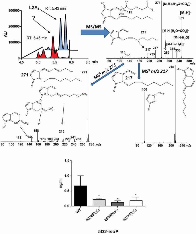
Figure 3.

Suggestive identification and measurement of fungal 5D2-IsoP in wild type and eicosanoid mutant strains. Upper left, LC/MS analysis of cell supernatant after growing C. parapsilosis wild type strain in presence of 100 μM arachidonic acid in PBS. Blue trace shows the elution of an authentic LXA4 standard (1 ng/mL) in the transition m/z 351-> 115. Red trace shows the elution of an overlapping signal in a representative C. parapsilosis sample. (Upper right) MS/MS spectrum of the signal co-eluting with LXA4 and chemical structure of the candidate molecule 5D2-IsoP. Middle MS3 spectra of m/z 271 (left) and m/z 217 (right). Lower panel, CPAR2_800020Δ/Δ showed a significant reduction in 5D2-IsoP production, although 5D2-IsoP levels also decreased in CPAR2_603600Δ/Δ and CPAR2_807710Δ/Δ strains.
