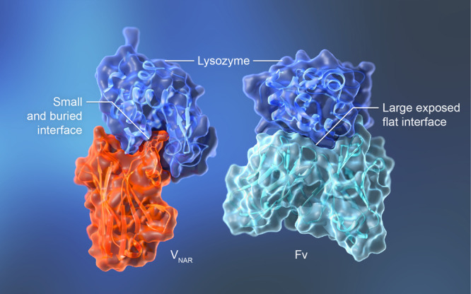
Figure 3.

The complex of a single-domain antibody and a protein antigen reveals a buried binding site. (a) The nurse shark VNAR single domain in complex with lysozyme (PDB 1T6V) [24]. VNAR residues Arg100 and Tyr101 are shown in the ribbon diagram to illustrate their interaction with the substrate binding pocket of lysozyme. (b) The humanized HyHEL-10 Fv in complex with lysozyme (PDB 2EIZ) [38].
