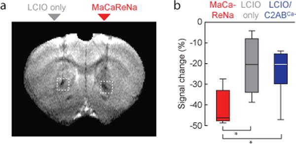
Figure 2. MaCaReNa calcium-binding activity correlates with MRI contrast in vivo.

(a) Injection of MaCaReNas into rat brain (right) induces substantially greater signal decrease than injection of LCIO nanoparticles alone (left). (b) Mean signal changes following intracranial injection of MaCaReNas (n = 7), LCIOs alone (n = 4), or calcium-insensitive LCIO/C2ABCa− mixtures (n = 5) were quantified in ROIs comparable to dotted white boxes in (a). Box plots indicate median (center line), first quartiles (box edges), and second quartiles (whiskers). Differences between MaCaReNas and both control conditions were significant (p ≤ 0.023).
