
Figure 1. Expression pattern of Pax6, FoxC1 and Pitx2 in the early and late eye.
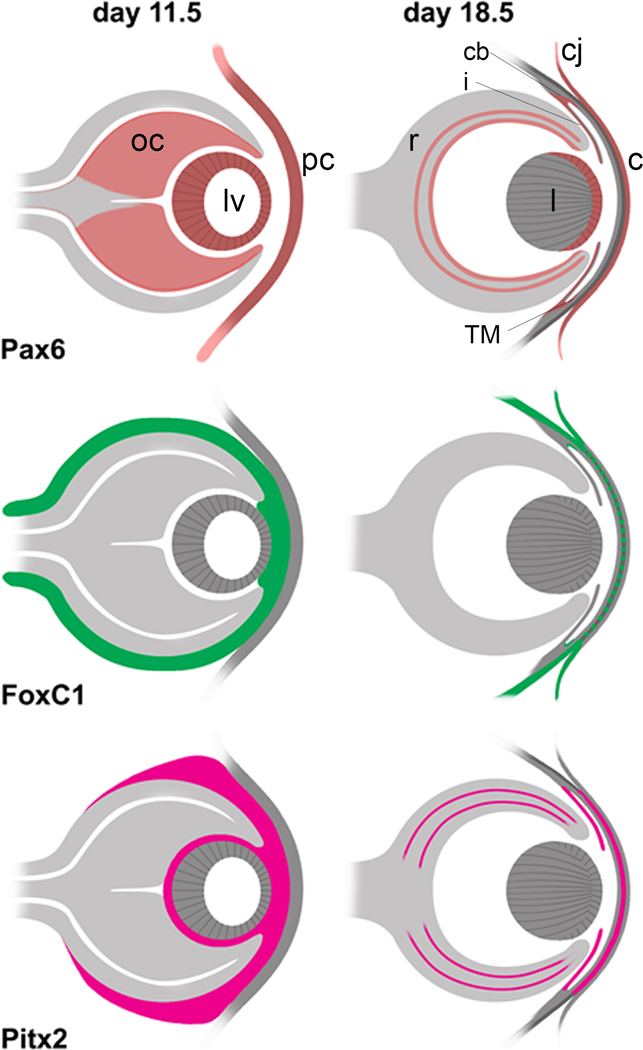
The expression patterns for Pax6, FoxC1, and Pitx2 are presented based on studies in mice and humans [82–87]. Early expression is shown at embryonic day 11.5 (E11.5), which represents day 33 in human development, and late expression is shown at E18.5, which is close to prenatal in humans. For Pax6, early expression is seen in the lens vesicle (lv), optic cup (oc), and presumptive cornea (pc) [86]. Expression later in the prenatal is restricted to the ganglion cell layer and inner and outer portions of the inner nuclear layer of the retina (r), where the latter points to expression specifically in the amacrine and horizontal cells [85]. At this stage, Pax6 is also expressed in the epithelia of the cornea (c), conjunctiva (cj), lens (l), ciliary body (cb), iris (i) and trabecular meshwork (TM). For both FoxC1 and Pitx2, expression in the early eye is seen in the neural crest cells of the periocular mesenchyme that surrounds the oc, in the intracellular space between the lv and pc, and the intracellular space between the inner oc and lv [84]. In the prenatal eye, FoxC1 expression is reduced to the conjunctival epithelium, sclera and trabecular meshwork [82] and Pitx2 expression is reduced to the iris [83], and according to a study of expression in human eyes is expressed in the corneal epithelium, the ciliary body non-pigmented layer and the nuclear layers of the retina [87]. Pitx2 is present in the corneal stroma but FoxC1 is much reduced in this layer [82, 83].